By Dr. Matthias Wiederholz with Performance Pain and Sports Medicine
Quadruple Board-Certified in Physical Medicine & Rehabilitation, Sports Medicine, Pain Medicine, and Regenerative Medicine
Updated January 23, 2026
Medically reviewed and updated for accuracy
Quick Insights: C5-C6 Bulging Disc Symptoms
What it is: A bulging disc at C5-C6 occurs when the intervertebral disc extends beyond its normal boundary, potentially compressing the C6 nerve root or spinal cord.
Common symptoms: Neck pain, radiating arm pain to the thumb and index finger, weakness in the biceps and wrist extensors, numbness, and tingling.
Who it affects: Most commonly develops in adults in midlife and beyond, though younger individuals with neck injuries or repetitive strain may also experience symptoms.
Treatment options: Range from conservative care (physical therapy, medications) to minimally invasive procedures like epidural injections and the Discseel® Procedure, with surgery reserved as a last resort.
Understanding C5-C6 Bulging Disc Symptoms
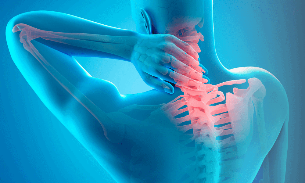
A bulging disc in the neck at the C5-C6 level represents one of the most common sources of cervical spine pain and neurological symptoms. This condition develops when the intervertebral disc between the fifth and sixth cervical vertebrae extends beyond its normal boundary, creating pressure on surrounding neural structures. The C5-C6 segment bears significant mechanical stress due to its location in the lower cervical spine, where the neck’s natural curve transitions and mobility demands are high.
Patients with a C5-C6 bulging disc typically present with a distinctive pattern of symptoms that reflect the specific nerve root affected. The C6 nerve root, which exits the spinal canal at this level, controls sensation and movement in specific areas of the upper extremity. Understanding these symptom patterns helps both patients and physicians identify the source of discomfort and develop appropriate treatment strategies. While many cases respond well to conservative interventions, recognizing when symptoms indicate more serious nerve compression or spinal cord involvement remains essential for optimal outcomes.
Cervical Spine Anatomy: The Foundation of Understanding
The cervical spine consists of seven vertebrae (C1-C7) that form the neck’s structural framework. Between each vertebra sits an intervertebral disc, a specialized structure composed of two distinct parts: a tough outer ring called the annulus fibrosus and a gel-like inner core known as the nucleus pulposus. These discs serve multiple critical functions—they act as shock absorbers during movement, allow for spinal flexibility, and maintain proper spacing between vertebrae to protect nerve roots as they exit the spinal canal.
The C5-C6 segment occupies a particularly vulnerable position in the lower cervical spine. This level experiences substantial mechanical loading during daily activities and bears considerable weight from the head and upper cervical spine. The natural lordotic curve of the cervical spine places additional stress on this segment, particularly during extension movements like looking upward. These biomechanical factors explain why C5-C6 ranks among the most common sites for disc degeneration and bulging in the cervical spine.
The C6 Nerve Root: Pathway and Function
The C6 nerve root emerges from the spinal canal through the neural foramen at the C5-C6 level. This nerve follows a specific pathway down the arm, providing both motor function to certain muscles and sensory perception to defined areas of skin. The C6 dermatome—the skin region supplied by this nerve—includes the lateral (outer) aspect of the forearm, the thumb, and the index finger. When a bulging disc compresses the C6 nerve root, patients typically experience symptoms in these specific distributions.
Motor functions controlled by the C6 nerve root include the biceps muscle (elbow flexion), the brachioradialis (forearm rotation), and the wrist extensor muscles. Compression or irritation of this nerve can lead to weakness in these muscle groups, manifesting as difficulty with specific tasks like lifting objects, turning doorknobs, or extending the wrist against resistance. Research published in StatPearls describes these precise neuroanatomical relationships and explains how disc pathology at C5-C6 produces the characteristic symptom patterns clinicians observe in practice.
Recognizing C5-C6 Bulging Disc Symptoms
The symptom presentation of a C5-C6 bulging disc follows predictable patterns based on the neuroanatomy of the C6 nerve root. However, symptom severity varies considerably between patients, ranging from mild intermittent discomfort to debilitating pain that significantly impacts daily function.
Pain Patterns and Characteristics
Neck pain represents the most common initial symptom of a C5-C6 bulging disc. This pain typically localizes to the lower cervical region but frequently radiates into the shoulder blade area and down the arm. The discomfort often intensifies with specific neck positions—particularly extension (looking upward) or rotation to the affected side. Many patients report that pain worsens at night or after prolonged periods of sitting, especially with poor posture that increases pressure on the anterior disc.
The radiating arm pain associated with C6 nerve root compression follows a characteristic distribution. Pain travels from the neck and shoulder down the lateral (thumb side) of the arm, into the forearm, and terminates in the thumb and index finger. This pain quality varies—some patients describe sharp, shooting sensations, while others experience a deep, aching discomfort. Coughing, sneezing, or straining can trigger sudden pain exacerbations due to temporary increases in spinal pressure.
Neurological Symptoms: Numbness, Tingling, and Weakness
Sensory disturbances accompany pain in most cases of C6 radiculopathy. Patients commonly report numbness or tingling sensations (paresthesias) in the thumb and index finger. These sensory changes may be constant or intermittent, often fluctuating based on neck position. Some individuals describe a feeling of the affected fingers being “asleep” or notice decreased ability to perceive fine touch or temperature differences in the C6 dermatome.
Motor weakness develops when nerve compression becomes more significant. C6 weakness most notably affects the biceps and wrist extensors. Patients may notice difficulty performing specific tasks: trouble lifting grocery bags (biceps weakness), inability to extend the wrist fully against resistance, or reduced grip strength. In clinical examination, physicians test these muscle groups systematically to confirm C6 nerve involvement. While imaging reveals the anatomical degree of nerve compression, research suggests that herniation size alone does not consistently predict the severity of motor deficits or clinical outcomes, as inflammatory mechanisms and individual factors often drive symptom severity more than compression visible on MRI.
Impact on Daily Activities and Range of Motion
A bulging disc at C5-C6 frequently restricts cervical spine mobility. Patients report stiffness when attempting to turn their head, particularly toward the affected side. This reduced range of motion stems from both mechanical factors (the disc bulge itself limiting movement) and protective muscle spasm that develops in response to pain. Activities requiring sustained neck positions become challenging—driving (especially checking blind spots), reading, or working at a computer often provoke symptoms.
The cumulative effect of these symptoms on quality of life can be substantial. Sleep disturbances are common, as finding a comfortable sleeping position becomes difficult. Work productivity may decline, particularly for individuals whose occupations involve computer use, overhead activities, or heavy lifting. Understanding these functional impacts helps guide treatment decisions and expectations for recovery timelines.
Degenerative Disc Disease at C5-C6
While the terms “bulging disc” and “degenerative disc disease” are sometimes used interchangeably, degenerative disc disease (DDD) represents a broader condition characterized by progressive changes in disc structure and function over time. At the C5-C6 level, DDD often underlies disc bulging and contributes to the development of symptoms.
The Degenerative Process: How Discs Age
Cervical disc degeneration follows a predictable sequence of changes that typically begin in the third or fourth decade of life. The process starts with loss of water content in the nucleus pulposus—the disc’s gel-like center—which reduces the disc’s ability to absorb shock and maintain proper height between vertebrae. As hydration decreases, the disc becomes less elastic and more prone to developing small tears in its outer annular fibers.
These structural changes create a cascade of effects throughout the motion segment. Disc height loss alters the biomechanics of the facet joints (small joints at the back of the spine), potentially leading to arthritis. The decreased disc space also narrows the neural foramen—the openings through which nerve roots exit—increasing the likelihood of nerve compression even without a significant disc bulge. Research in StatPearls on cervical degenerative disc disease describes these pathophysiological changes and notes that the C5-C6 level experiences particularly high rates of degeneration due to its biomechanical loading patterns.
Symptoms Specific to C5-C6 Degenerative Disc Disease
Degenerative disc disease at C5-C6 produces symptoms that may be more insidious in onset compared to acute disc injuries. Many patients experience gradually worsening neck stiffness and discomfort that develops over months or years. Morning stiffness that improves with movement represents a hallmark feature. The pain quality often differs from acute radiculopathy—patients describe deep, aching neck pain that may not radiate as distinctly down the arm initially.
As degeneration progresses, nerve root compression becomes more likely due to a combination of factors: disc space narrowing, facet joint hypertrophy (enlargement), and occasional disc bulging or herniation. When C6 radiculopathy develops in the context of DDD, symptoms may fluctuate more than with an acute disc herniation. Patients often identify positions or activities that consistently provoke symptoms, suggesting mechanical compression that varies with neck position. For a more comprehensive understanding of cervical degenerative disc disease and its progression through different cervical levels, additional resources provide detailed insights into long-term management strategies.
Radiographic Findings: Disc Space Narrowing
Imaging studies reveal characteristic findings in cervical DDD. Plain X-rays demonstrate disc space narrowing at C5-C6—a reduction in the normal height between these vertebrae. Advanced imaging with MRI provides more detailed information about disc hydration, annular tears, and the degree of neural compression. The Pfirrmann classification system, commonly used to grade disc degeneration, categorizes severity from Grade I (normal disc with bright signal on MRI) to Grade V (severe degeneration with collapsed disc space and dark signal).
Clinical observation and research on asymptomatic adults demonstrates that imaging findings don’t always correlate perfectly with symptom severity. Studies of asymptomatic adults show a high prevalence of cervical disc abnormalities on MRI—including 81% with disc herniation, 85.9% with annular fissure, and 95.4% with nucleus degeneration—underscoring that some patients with advanced radiographic degeneration report minimal symptoms, while others with moderate imaging changes experience significant disability. This observation emphasizes the importance of treating patients based on their clinical presentation rather than imaging alone.
Disc Space Narrowing at C5-C6: Clinical Significance
Disc space narrowing at the C5-C6 level represents both a radiographic finding and a mechanical problem with real clinical consequences. As the vertical height between vertebrae decreases, multiple structures experience altered biomechanics and increased stress.
Mechanisms and Contributing Factors
Disc space narrowing develops through several pathways. Chronic degeneration represents the most common cause, but acute disc injuries can also produce rapid height loss if nuclear material extrudes. Repetitive microtrauma from occupational activities or sports participation may accelerate the process. Genetic factors influence individual susceptibility to disc degeneration—some families demonstrate earlier and more severe disc disease than the general population.
The consequences of disc space narrowing extend beyond simple height loss. As vertebrae move closer together, the neural foramen narrows, potentially trapping nerve roots even without a disc bulge. The uncovertebral joints (small joints on the sides of cervical vertebrae) experience increased loading and may develop bone spurs (osteophytes) that further compromise neural space. These secondary changes often contribute as much to symptoms as the original disc pathology.
Symptoms Associated with Disc Space Narrowing
Patients with significant disc space narrowing at C5-C6 often report symptoms that worsen gradually over time rather than beginning with a specific injury. Chronic neck pain and stiffness predominate. Nerve root symptoms may be positional—certain head positions temporarily increase foraminal narrowing and provoke arm pain or numbness. Morning symptoms often prove worse, as overnight disc hydration slightly increases disc height, which then decreases throughout the day with normal activity.
When disc space narrowing becomes severe, instability may develop at the affected segment. Patients describe a sensation of the neck “giving way” or feeling unstable during certain movements. Abnormal motion between vertebrae can produce intermittent nerve compression, leading to unpredictable symptom patterns that seem disproportionate to the imaging findings.
Treatment Considerations for Disc Space Narrowing
Management of symptomatic disc space narrowing requires a comprehensive approach. Conservative treatments focus on maintaining flexibility, strengthening supporting muscles, and modifying activities that exacerbate symptoms. Cervical traction may temporarily increase disc space and provide symptom relief, though effects are typically temporary. Anti-inflammatory medications help manage pain but don’t alter the underlying structural problem.
For patients with persistent symptoms despite conservative management, interventional procedures offer additional options. Epidural steroid injections can reduce nerve root inflammation and provide intermediate-term relief. The Discseel® Procedure addresses disc pathology directly by sealing annular tears with a biologic agent, potentially stabilizing the disc and preventing further height loss. When disc space narrowing leads to severe instability or progressive neurological deficits, surgical options including fusion or disc replacement may be considered.
Distinguishing Radiculopathy from Myelopathy at C5-C6
One of the most critical distinctions in evaluating C5-C6 disc pathology involves differentiating cervical radiculopathy from cervical myelopathy. While both conditions can result from disc-related compression, they involve different neural structures and carry different implications for treatment urgency and prognosis.
Cervical Radiculopathy: Nerve Root Compression
Cervical radiculopathy occurs when a bulging or herniated disc compresses a nerve root—in this case, the C6 nerve root as it exits the spinal canal. Symptoms follow the dermatomal and myotomal distribution of the affected nerve. As described earlier, C6 radiculopathy produces pain, numbness, and tingling in the lateral arm, thumb, and index finger, along with potential weakness in the biceps and wrist extensors.
Radiculopathy symptoms typically remain unilateral (affecting one arm) unless bilateral disc herniations occur, which is uncommon. The pain often has a sharp or burning quality and may be provoked by specific neck positions that increase nerve compression. Spurling’s test—a clinical maneuver where the examiner extends and rotates the neck toward the symptomatic side while applying downward pressure—often reproduces radicular symptoms when the C6 nerve root is compressed.
Cervical Myelopathy: Spinal Cord Compression
Cervical myelopathy represents a more serious condition where the spinal cord itself experiences compression. At C5-C6, a large central disc herniation or severe disc bulge combined with pre-existing spinal canal narrowing can compress the spinal cord. According to Cleveland Clinic’s comprehensive review of cervical myelopathy, symptoms differ significantly from radiculopathy and include:
- Bilateral symptoms: Both arms and potentially both legs may be affected, as the spinal cord carries nerve fibers to all four extremities
- Hand clumsiness: Difficulty with fine motor tasks like buttoning shirts or handling small objects
- Gait disturbances: Unsteady walking, balance problems, or difficulty with stairs
- Bowel or bladder dysfunction: In severe cases, loss of normal urinary or bowel control
- Hyperreflexia: Abnormally brisk reflexes in the arms and legs, often with positive Hoffman’s sign or Babinski reflex
The American Academy of Orthopaedic Surgeons emphasizes that cervical myelopathy typically develops gradually but can progress to permanent spinal cord damage if left untreated. Unlike radiculopathy, which often improves with conservative treatment, myelopathy usually requires surgical decompression to prevent neurological deterioration.
Why the Distinction Matters
Correctly identifying whether symptoms stem from radiculopathy or myelopathy profoundly impacts treatment decisions. Radiculopathy can often be managed conservatively with physical therapy, medications, and potentially minimally invasive procedures. Many patients experience significant improvement within weeks to months. Myelopathy, conversely, represents a more urgent condition that typically requires surgical intervention to decompress the spinal cord and prevent permanent neurological injury.
In clinical practice, some patients present with mixed pictures—radiculopathy symptoms with subtle myelopathic signs. MRI imaging becomes essential in these cases to visualize the degree and location of neural compression. The presence of spinal cord signal changes on MRI (indicating cord injury) warrants prompt neurosurgical or spine surgery consultation.
Diagnostic Approach to C5-C6 Disc Pathology
Accurate diagnosis of C5-C6 bulging disc and related conditions requires a systematic approach combining clinical evaluation and imaging studies. The diagnostic process aims to confirm the anatomical source of symptoms, assess severity, and identify any features requiring urgent intervention.
Clinical Examination
The physical examination for suspected C5-C6 disc pathology includes several components. Inspection may reveal loss of normal cervical lordosis or protective muscle guarding. Range of motion testing documents limitations in cervical flexion, extension, and rotation. Provocative maneuvers like Spurling’s test help localize nerve root compression, while the shoulder abduction relief test (raising the arm overhead to reduce symptoms) suggests radiculopathy.
Neurological examination systematically assesses motor function, sensation, and reflexes. For C6 radiculopathy, physicians test biceps strength, wrist extension strength, and the brachioradialis reflex. Sensory examination maps any deficits in the C6 dermatome. When myelopathy is suspected, additional testing includes assessment of hand coordination, gait evaluation, and testing for pathological reflexes that indicate spinal cord involvement.
Imaging Studies
MRI represents the gold standard for evaluating cervical disc pathology. This imaging modality visualizes soft tissue structures including discs, nerve roots, and the spinal cord with excellent detail. MRI can differentiate disc bulges from herniations, assess disc hydration and degeneration severity, and identify nerve root or spinal cord compression. Signal changes within the spinal cord itself appear on MRI when myelopathy is present, providing important prognostic information.
Plain radiographs (X-rays), while less detailed than MRI, offer valuable information about overall spinal alignment, disc space height, and the presence of bone spurs or vertebral instability. Flexion-extension X-rays can reveal abnormal motion between vertebrae. CT scanning provides excellent bone detail and may be ordered when MRI is contraindicated or when surgical planning requires precise bone anatomy mapping.
Electrodiagnostic Testing
Electromyography (EMG) and nerve conduction studies help confirm the diagnosis of radiculopathy and distinguish it from other conditions affecting the upper extremity. These tests measure electrical activity in muscles and nerves, identifying patterns consistent with C6 nerve root compression. While not always necessary, electrodiagnostic testing proves particularly useful when clinical and imaging findings don’t clearly correlate or when peripheral nerve conditions might explain symptoms.
Treatment Strategies for C5-C6 Bulging Disc
Management of C5-C6 bulging disc symptoms follows a stepwise approach, beginning with conservative measures and progressing to more invasive interventions only when necessary. Systematic reviews of conservative treatment for cervical radiculopathy indicate that patients tend to improve over time with a generally favorable natural course, though individual results vary based on multiple factors.
Conservative Treatment Options
Physical therapy forms the cornerstone of conservative management. A skilled physical therapist designs exercises to strengthen cervical and periscapular muscles, improve posture, and restore normal neck mechanics. Manual therapy techniques including gentle mobilization may provide pain relief and improve range of motion. Education about proper ergonomics during work and daily activities helps prevent symptom exacerbation.
Medications play an adjunctive role in symptom control. Non-steroidal anti-inflammatory drugs (NSAIDs) reduce inflammation around compressed nerve roots and alleviate pain. Neuropathic pain medications such as gabapentin or pregabalin may help when nerve-related symptoms predominate. Muscle relaxants address protective muscle spasm that often accompanies disc pathology. For patients unable to tolerate oral medications, topical treatments including prescription lidocaine patches deliver medication directly to affected areas.
Cervical traction devices provide temporary symptom relief for some patients by gently separating vertebrae and reducing nerve root compression. Activity modification—avoiding positions and movements that provoke symptoms—allows inflammation to subside. Many patients benefit from a brief period of relative rest followed by gradual return to normal activities as symptoms improve.
Minimally Invasive Interventional Procedures
When conservative treatments fail to provide adequate relief, minimally invasive procedures offer effective alternatives to surgery. Epidural steroid injections deliver potent anti-inflammatory medication directly to the site of nerve root compression. These injections can provide significant pain relief lasting weeks to months, allowing patients to participate more effectively in physical therapy and potentially avoiding surgery altogether.
The Discseel® Procedure represents an innovative approach to treating disc pathology at its source. This minimally invasive treatment involves injecting a biologic fibrin sealant into the disc to seal annular tears and stabilize the disc structure. Dr. Matthias Wiederholz, the first physician in Houston trained to perform the Discseel® Procedure, received direct instruction from the technique’s inventor, Dr. Kevin Pauza. Dr. Wiederholz was the first physician in Houston trained to perform the Discseel® Procedure and has been offering this treatment since 2020, establishing extensive experience as Houston’s pioneer in this approach.
The Discseel® Procedure offers several advantages for appropriate candidates. Unlike surgery, it preserves the disc and doesn’t require hardware implantation or bone fusion. The procedure is performed on an outpatient basis with minimal recovery time compared to surgical interventions. By sealing disc tears, the treatment addresses the underlying pathology rather than simply managing symptoms. Patients seeking to avoid the risks and lengthy recovery associated with cervical fusion surgery often find the Discseel® Procedure an attractive option.
The Discseel® Procedure at Performance Pain and Sports Medicine
Dr. Wiederholz’s expertise with the Discseel® Procedure stems from his direct training under Dr. Kevin Pauza, the inventor of this treatment. This firsthand instruction ensures that patients receive the highest standard of care using the most current techniques. As Houston’s pioneer in offering the Discseel® Procedure and one of only three Master Instructors worldwide, Dr. Wiederholz has helped numerous patients find relief from chronic neck pain without resorting to surgery.
The procedure involves precise, fluoroscopy-guided injection of fibrin into damaged discs. This biologic agent seals annular tears, preventing further disc degeneration and reducing inflammation around nerve roots. Many patients experience gradual symptom improvement over the months following treatment as the disc heals and stabilizes. Individual results vary.
If you’re considering the Discseel® Procedure for C5-C6 disc pathology, consultation with Dr. Wiederholz includes comprehensive evaluation, review of imaging studies, and discussion of expected outcomes based on your specific condition. Learn more about the Discseel® Procedure and whether you might be a candidate for this treatment.
Surgical Interventions
Surgery becomes necessary when conservative and minimally invasive treatments fail to provide relief, when progressive neurological deficits develop, or when myelopathy is present. Several surgical options exist for C5-C6 disc pathology, each with specific indications and expected outcomes.
Anterior cervical discectomy and fusion (ACDF) remains the most common surgical procedure for symptomatic C5-C6 disc disease. The surgeon removes the damaged disc through an incision in the front of the neck, decompresses the nerve root or spinal cord, and then fuses the C5 and C6 vertebrae together using a bone graft and metal hardware. A 2024 systematic review comparing ACDF with cervical disc arthroplasty found that ACDF provides reliable long-term outcomes for cervical radiculopathy and myelopathy, though it eliminates motion at the treated level.
Cervical disc replacement (arthroplasty) offers an alternative that preserves motion at the C5-C6 level. This procedure involves removing the damaged disc and replacing it with an artificial disc prosthesis. Potential advantages include maintained neck mobility and reduced stress on adjacent spinal levels. However, not all patients are candidates for disc replacement—factors like significant arthritis or osteoporosis may favor fusion instead.
Posterior cervical foraminotomy represents a less common but sometimes appropriate surgical approach. This procedure enlarges the neural foramen from the back of the neck, decompressing the nerve root without removing the disc or performing a fusion. Johns Hopkins Medicine notes that foraminotomy works best for patients with primarily posterolateral disc herniations and may preserve more normal neck motion than anterior approaches.
Prevention and Long-Term Management
While some risk factors for C5-C6 disc degeneration (age, genetics) cannot be modified, several strategies can reduce symptom severity and potentially slow progression of disc disease.
Ergonomics and Posture
Proper posture during work and daily activities significantly impacts cervical spine health. Forward head posture—common with prolonged computer use or smartphone viewing—dramatically increases stress on the lower cervical spine. For every inch the head moves forward from ideal alignment, the effective weight on the cervical spine increases substantially. Maintaining the head directly over the shoulders, using properly positioned computer monitors, and taking frequent breaks from sustained postures all help reduce C5-C6 loading.
Sleeping position matters as well. Pillows should support the natural cervical curve without forcing the neck into extreme flexion or extension. Side sleepers benefit from pillows that maintain the head in neutral alignment with the spine, while back sleepers typically need thinner pillows to avoid excessive neck flexion.
Exercise and Strengthening
Regular exercise targeting the neck, upper back, and shoulder blade muscles provides dynamic stability for the cervical spine. Chin tucks strengthen deep neck flexor muscles. Scapular retraction exercises combat rounded shoulder posture. Gentle cervical range of motion exercises maintain flexibility. A physical therapist can design a personalized program appropriate for individual needs and limitations.
General cardiovascular fitness and core strengthening indirectly benefit the cervical spine by improving overall posture and reducing compensation patterns that increase neck stress. Maintaining healthy body weight reduces mechanical loading on all spinal segments.
Activity Modification
Certain activities place particularly high stress on the C5-C6 segment and may need modification. Overhead work, heavy lifting, and contact sports all increase risk of disc injury. While these activities don’t always need to be eliminated, proper technique and gradual progression help minimize injury risk. Patients with known C5-C6 disc pathology benefit from discussing activity restrictions and modifications with their treating physician.
Frequently Asked Questions
1. What triggers C5-C6 bulging disc symptoms?
Certain positions and movements commonly provoke symptoms. Looking upward (cervical extension) narrows the neural foramen and can increase nerve compression. Rotation toward the affected side often reproduces radicular pain. Heavy lifting, prolonged sitting with poor posture, and activities involving repetitive neck movements may trigger symptom flares. Many patients report worse pain at night or upon waking, possibly due to prolonged static postures during sleep.
2. How can I tell if I have radiculopathy or myelopathy?
Radiculopathy typically causes unilateral arm symptoms (pain, numbness, weakness) following a specific nerve root distribution. C6 radiculopathy affects the thumb side of the arm and hand. Myelopathy involves the spinal cord and produces bilateral symptoms, hand clumsiness, gait difficulties, and potential bowel or bladder changes. Myelopathy represents a more serious condition requiring urgent evaluation. If you experience symptoms in both arms, balance problems, or difficulty with fine hand movements, seek prompt medical attention.
3. Can C5-C6 bulging disc symptoms resolve without surgery?
Many patients with C5-C6 radiculopathy improve with conservative treatment. Studies show that patients with cervical radiculopathy tend to improve over time with a generally favorable natural course, though individual outcomes vary. Physical therapy, medications, activity modification, and sometimes epidural injections provide relief for most individuals. However, patients with severe or progressive weakness, myelopathy, or symptoms persisting beyond three months despite conservative treatment may benefit from surgical consultation.
4. What is the Discseel® Procedure and am I a candidate?
The Discseel® Procedure is a minimally invasive treatment that seals disc tears using a biologic fibrin agent. Ideal candidates have symptomatic disc pathology confirmed on MRI, persistent symptoms despite conservative treatment, but have not yet developed severe spinal cord compression requiring urgent decompression. During consultation, our team reviews your imaging studies and clinical presentation to determine if the Discseel® Procedure might benefit you. Not all patients are candidates, but for appropriate individuals, this treatment offers an alternative to surgery with potentially excellent results.
5. How long does recovery take after treatment?
Recovery timelines vary by treatment modality. Conservative treatment typically shows improvement within weeks to months. Epidural injections may provide relief within days to a week, though full benefits sometimes take longer. The Discseel® Procedure involves gradual healing over three to six months as the disc stabilizes and inflammation resolves. Surgical recovery depends on the specific procedure—minimally invasive approaches may allow return to light activities within weeks, while fusion surgery typically requires three to six months for bone healing and return to full activity.
6. Can disc bulging at C5-C6 recur after treatment?
Recurrence risk depends on multiple factors including the treatment received, adherence to preventive strategies, and ongoing disc degeneration. After successful conservative treatment or epidural injections, symptoms may recur if the underlying disc pathology persists. The Discseel® Procedure aims to stabilize the disc and reduce recurrence risk by sealing tears. Following surgical fusion, the treated level cannot develop recurrent symptoms, though adjacent segments may experience increased stress over time. Maintaining good posture, appropriate exercise, and activity modifications help minimize recurrence risk regardless of treatment modality.

Conclusion
A bulging disc at C5-C6 produces a characteristic pattern of symptoms reflecting the anatomy of the C6 nerve root and the biomechanical stress placed on this vulnerable spinal segment. While neck pain and radiating arm discomfort can significantly impact quality of life, understanding the condition empowers patients to make informed treatment decisions. The distinction between radiculopathy and myelopathy proves crucial, as these conditions require different management approaches.
Treatment options span a broad spectrum from conservative care to advanced surgical interventions. Many patients achieve excellent outcomes with physical therapy and medications alone. For those requiring additional intervention, minimally invasive procedures like epidural injections and the Discseel® Procedure offer effective alternatives to surgery. When surgery becomes necessary, modern techniques provide reliable results for carefully selected candidates.
Prevention and long-term management strategies—proper ergonomics, regular exercise, and activity modification—help minimize symptom recurrence and potentially slow progressive disc degeneration. By combining evidence-based treatments with proactive self-care, patients with C5-C6 bulging discs can often return to comfortable, active lifestyles.
If you’re experiencing neck pain, arm symptoms, or other signs of C5-C6 disc pathology, Performance Pain and Sports Medicine offers comprehensive evaluation and treatment. Dr. Matthias Wiederholz provides expert care including the Discseel® Procedure at our Houston, Texas location. Our team develops personalized treatment plans designed to address your specific needs and help you return to the activities you enjoy. Schedule a consultation to explore your treatment options.
Medical Disclaimer
This article is for informational and educational purposes only and does not constitute medical advice. The information provided should not be used for diagnosing or treating a health condition. Always consult with a qualified healthcare provider regarding any medical concerns, symptoms, or treatment options. Individual results may vary, and treatment recommendations depend on each patient’s unique clinical presentation. Performance Pain and Sports Medicine provides this content to help patients make informed decisions in consultation with their physicians.