By Dr. Matthias Wiederholz with Performance Pain and Sports Medicine
Quadruple Board-Certified in Physical Medicine & Rehabilitation, Sports Medicine, Pain Medicine, and Regenerative Medicine
Updated February 12, 2026
Medically reviewed and updated for accuracy
Quick Insights
A herniated disc x ray cannot directly show the disc itself because X-rays capture bone, not soft tissue. However, X-rays may reveal indirect signs of disc problems, including disc space narrowing, bone spurs, and vertebral misalignment. For a definitive diagnosis, MRI remains the gold standard imaging tool. Whether you call it a herniated disc, slipped disc, or bulging disc, the same imaging limitations apply. If you suspect a disc injury, understanding what each test can and cannot show helps you take the right next step toward relief.
Key Takeaways
- X-rays cannot visualize herniated or slipped discs directly because discs are soft tissue structures invisible on plain radiographs.
- X-rays can reveal indirect signs such as disc space narrowing, endplate sclerosis, and osteophyte formation that suggest underlying disc degeneration.
- MRI is the preferred diagnostic tool, with pooled sensitivity of 89% and specificity of 83% for lumbar disc herniation.
- Cervical spine X-rays have the same soft tissue limitations, making MRI essential for diagnosing neck disc herniations.
- Over 85% of acute herniated disc symptoms resolve within 8 weeks with conservative management, and imaging is typically recommended only when symptoms persist or red flags are present.
Why It Matters
Many patients with back or neck pain assume an X-ray will reveal whether they have a herniated disc. When results come back “normal,” they may feel confused or dismissed. Understanding that X-rays simply cannot capture soft tissue disc pathology empowers you to have an informed conversation with your physician about next steps. The right imaging at the right time leads to accurate diagnosis, and accurate diagnosis leads to effective treatment.
Introduction
As an interventional spine specialist in Houston, one of the most common questions I hear from patients is whether a herniated disc x ray can confirm their diagnosis. The straightforward answer is that X-rays alone cannot directly show a herniated disc. X-rays are excellent at capturing images of bones and bony structures, but the intervertebral discs that cushion your spine are soft tissue, and soft tissue does not appear on standard radiographs.
That said, X-rays still play a valuable role in the diagnostic process. They can reveal indirect clues, such as narrowing between vertebrae or bone spur formation, that suggest disc problems may be present. These findings often guide physicians toward ordering more detailed imaging like MRI to confirm the diagnosis. Dr. Matthias Wiederholz and the team at Performance Pain and Sports Medicine use a comprehensive diagnostic approach to identify the true source of your pain and develop an individualized treatment plan.
This article walks you through what X-rays can and cannot show, when more advanced imaging is needed, and what treatment options are available once a herniated disc is confirmed. Whether you have been told your X-ray looks “normal” or you are trying to understand results that mention disc space narrowing, this guide is for you.
What Is a Herniated Disc?
Anatomy of the Spine and Discs
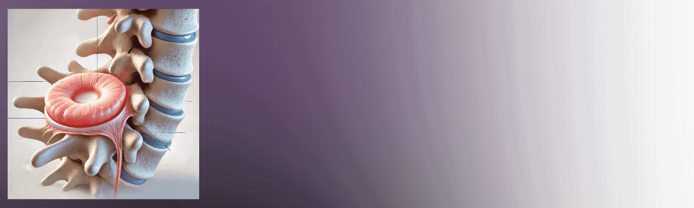
The human spine consists of 33 vertebrae stacked in a column, separated by intervertebral discs that act as shock absorbers. Each disc has two main components: a tough outer ring called the annulus fibrosus and a soft, gel-like center known as the nucleus pulposus. Together, these structures allow the spine to flex, extend, and rotate while distributing mechanical forces during daily activities.
How Herniated Discs Develop
A herniated disc occurs when the nucleus pulposus pushes through a weakened area or tear in the annulus fibrosus. This can happen gradually through age-related wear and tear, or suddenly from an injury such as a fall, lifting accident, or car collision. Over 3 million people in the United States are diagnosed with a herniated disc each year, and the condition goes by several names, including slipped disc, bulging disc, ruptured disc, and protruding disc. Regardless of the terminology, the underlying mechanism is the same: disc material extends beyond its normal boundary and may compress nearby spinal nerves.
Common Symptoms
Symptoms depend on where the herniation occurs and whether it compresses a nerve root. Patients commonly experience localized back pain or neck pain, radiating pain into the arms or legs, numbness and tingling along a nerve pathway, and muscle weakness affecting grip strength, balance, or walking. Some herniations produce minimal symptoms, while others cause significant functional limitations that interfere with work, sleep, and daily life.
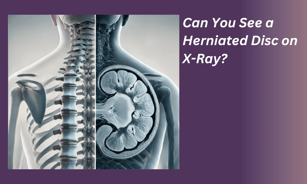
Can a Herniated Disc Be Seen on an X-Ray?
The short answer is no. A standard X-ray cannot directly visualize a herniated disc. X-rays work by passing electromagnetic radiation through the body, where dense structures like bone absorb more radiation and appear white on the image. Soft tissues, including intervertebral discs, spinal nerves, and the spinal cord, do not absorb enough radiation to create a distinct image. This means the disc itself is essentially invisible on a plain radiograph.
The World Federation of Neurosurgical Societies (WFNS) Spine Committee published consensus recommendations in 2024 confirming that MRI is the gold standard for diagnosing lumbar disc herniation. The committee noted that plain X-ray images can be considered only as an adjunct imaging modality for differential diagnosis, not as a primary diagnostic tool for disc pathology.
So if X-rays cannot show the disc, why do physicians still order them? Because they provide important information about bony alignment, vertebral fractures, and structural changes that may be contributing to your symptoms. X-rays are often the first step in a diagnostic process that may lead to more detailed imaging when needed.

What Can an X-Ray Reveal in Suspected Disc Problems?
While X-rays cannot show a herniated disc directly, they can reveal several indirect signs that point toward underlying disc pathology. Research on degenerative disc disease radiographic findings identifies the key features visible on plain radiographs:
Disc space narrowing is one of the most significant X-ray findings. When a disc loses hydration and height over time, the space between adjacent vertebrae shrinks. This reduced disc height is measurable on X-ray and suggests that the disc has undergone degeneration, a process that increases the likelihood of herniation.
Endplate sclerosis appears as increased whiteness (density) along the edges of the vertebral bodies where they contact the disc. This hardening of bone occurs as a response to abnormal mechanical stress from a deteriorating disc.
Osteophyte formation refers to bone spurs that develop along the vertebral margins. These bony projections grow as the body attempts to stabilize a segment where disc support has weakened. Osteophytes can sometimes contribute to nerve compression on their own.
Vertebral misalignment or instability may also be visible, particularly on flexion-extension X-rays taken while you bend forward and backward. These dynamic views can reveal abnormal movement between vertebrae that suggests the disc is no longer providing adequate stability.
When any of these findings appear, your physician will typically recommend further evaluation with MRI to determine the actual condition of the disc and surrounding soft tissues.
Can You See a Slipped Disc on an X-Ray?
No, you cannot see a slipped disc on an X-ray for the same reasons you cannot see a herniated disc. “Slipped disc” is a widely used term, particularly in the United Kingdom and internationally, that describes the same condition as a herniated disc. The Mayo Clinic confirms that plain X-rays do not detect herniated disks, though they can help rule out other causes of back pain such as infection, tumor, spinal alignment problems, or a broken bone.
The terminology can be confusing. Whether your physician, physical therapist, or imaging report uses the term herniated disc, slipped disc, bulging disc, or ruptured disc, the X-ray limitations remain identical. The disc material is soft tissue and will not appear on a standard radiograph regardless of which name is used.
If you have been told your slipped disc X-ray looks “normal,” that does not necessarily mean nothing is wrong. It simply means that X-ray is not the right tool to evaluate disc pathology. Many patients with significant disc herniations have completely normal-appearing X-rays because the bony structures may look fine even when the disc between them is damaged.

Can You See Degenerative Disc Disease on an X-Ray?
Unlike a herniated disc, some features of degenerative disc disease (DDD) can be identified on X-ray. DDD develops when intervertebral discs gradually lose hydration, height, and structural integrity over time. Because this process produces measurable changes in the bony structures surrounding the disc, X-rays can detect several hallmarks of degeneration.
The most recognizable X-ray finding in DDD is reduced disc height, where the space between two vertebrae appears narrower than normal. Additional findings include endplate irregularities, vacuum phenomena (small pockets of gas within a degenerating disc), and osteophyte formation along the vertebral margins. These radiographic signs help physicians gauge how much degeneration has occurred at a given spinal level.
However, X-ray alone cannot tell the full story. While it may show that a disc has lost height, it cannot reveal whether that disc is also herniated, whether there is nerve compression, or whether the disc contains annular tears that may be generating pain. The North American Spine Society (NASS) clinical guidelines recommend MRI as the most appropriate noninvasive test when clinical history and physical examination suggest disc herniation with radiculopathy, because MRI can visualize the soft tissue changes that X-ray cannot.
Herniated Disc in Neck on X-Ray: Cervical Spine Limitations
The same imaging limitations that apply to lumbar (lower back) disc herniations also apply to the cervical (neck) spine. A herniated disc in the neck will not appear on an X-ray. Cervical spine X-rays can show the alignment of your neck vertebrae, the presence of bone spurs, and whether disc spaces have narrowed, but they cannot visualize the disc material itself or confirm whether it is compressing a cervical nerve root or the spinal cord.
Research on cervical radiculopathy has demonstrated that radiographic findings alone may be inadequate for diagnosing cervical disc problems, and that additional testing such as electromyography (EMG) adds diagnostic value beyond what plain films and clinical examination provide. Cervical disc herniations can cause arm pain, hand numbness, grip weakness, and in severe cases, difficulty with coordination and balance.
When a cervical herniation is suspected based on your symptoms and physical examination, cervical MRI is the imaging study of choice. It provides detailed views of the discs, nerve roots, and spinal cord that are simply not possible with X-ray.

Imaging Tests Beyond X-Rays for Herniated Discs
MRI: The Gold Standard
MRI (magnetic resonance imaging) is universally recognized as the preferred diagnostic tool for herniated discs. Unlike X-rays, MRI uses magnetic fields and radio waves to create highly detailed images of soft tissues, including intervertebral discs, nerve roots, and the spinal cord. A 2023 systematic review and meta-analysis published in Frontiers in Surgery found that MRI has a pooled sensitivity of 89% and specificity of 83% for diagnosing lumbar disc herniation, based on 38 studies involving 1,875 patients.
MRI can identify the exact location and size of a herniation, show whether nerve roots are being compressed, and detect associated findings like annular tears, disc dehydration, and spinal canal narrowing. This level of detail is essential for determining the most appropriate treatment approach.
CT Scans
CT (computed tomography) scans combine multiple X-ray images to create cross-sectional views of the spine. They provide excellent visualization of bony structures and can detect disc herniations in some cases, particularly when paired with contrast dye (CT myelography). CT may be recommended for patients who cannot undergo MRI due to metal implants or other contraindications.
CT Myelography
For complex cases, a myelogram with CT scanning may be recommended. This involves injecting contrast dye into the spinal canal to highlight the spinal cord and nerve roots. The subsequent CT scan can reveal nerve root compression and structural issues that may not be clearly visible on standard imaging. This technique is particularly useful when MRI results are inconclusive or when surgical planning requires additional detail.
Nerve Tests to Assess Herniated Disc Impact
Electromyography (EMG)
EMG testing measures electrical activity in muscles to detect nerve damage or compression caused by a herniated disc. By identifying which muscles show abnormal electrical patterns, EMG helps pinpoint the specific nerve root being affected. This functional information complements the structural information provided by imaging.
Nerve Conduction Studies (NCS)
Nerve conduction studies, often performed alongside EMG, measure the speed and strength of electrical signals traveling along specific nerve pathways. Research confirms that EMG and imaging provide complementary information in radiculopathy, with electrophysiology adding diagnostic clarity that imaging alone may not provide. Together, these tests help your physician understand not just where a disc herniation exists, but how significantly it is affecting nerve function.

The Diagnostic Process: What to Expect
Medical History and Physical Examination
The diagnostic process begins with a thorough medical history and physical examination. Your physician will ask about the onset, location, and severity of your symptoms, what activities make them better or worse, and whether you have experienced any neurological changes such as numbness, tingling, or weakness.
Neurological Evaluation
A comprehensive neurological exam tests your reflexes, sensation, and muscle strength to identify areas of nerve compression. Specific physical examination maneuvers, such as the straight leg raise test for lumbar radiculopathy, can help localize which nerve root may be involved.
When Imaging Is Ordered
Current clinical guidelines indicate that imaging studies are not recommended in patients without red flag signs unless symptoms persist beyond 6 weeks. Red flags that warrant earlier imaging include progressive neurological deficits, suspected cauda equina syndrome, history of cancer, unexplained weight loss, fever, or recent significant trauma. This approach avoids unnecessary radiation exposure and ensures imaging is used when it will most meaningfully guide treatment decisions. The American College of Radiology Appropriateness Criteria reinforce that MRI is preferred over X-ray when interventional treatment planning is being considered.

Treatment Options After Diagnosis
Conservative Management
Most patients with a confirmed herniated disc begin with conservative treatment. Research shows that over 85% of acute herniated disc symptoms resolve within 8 weeks with appropriate conservative management. Treatment may include physical therapy to strengthen core and spinal stabilizer muscles, anti-inflammatory medications to reduce swelling around compressed nerves, activity modification to avoid aggravating positions, and epidural steroid injections for targeted pain relief when oral medications are insufficient.
Surgical Options for Severe Cases
If conservative treatment fails to provide adequate relief after a reasonable trial period, surgical options may be considered. Procedures such as microdiscectomy remove the portion of disc material compressing a nerve root. Spinal fusion may be recommended in cases where structural instability is present. However, surgery is typically reserved as a last resort after other approaches have been thoroughly explored.
The Discseel® Procedure: A Minimally Invasive Alternative
For patients seeking an alternative to traditional surgery, the Discseel® Procedure offers a regenerative approach to disc repair. This minimally invasive treatment uses a biologic fibrin sealant to repair annular tears in damaged discs, addressing the structural root cause of discogenic pain rather than just managing symptoms.
Peer-reviewed outcomes data published in Pain Physician in 2024 demonstrated statistically significant improvement across all outcome measures at 3, 6, 12, 24, and 36 months following treatment. The 320-patient study reported no adverse events and documented sustained benefit even in patients who had failed all prior treatments. Dr. Wiederholz was the first physician in Houston trained to perform the Discseel® Procedure, having received direct instruction from the procedure’s inventor, Dr. Kevin Pauza.
If you are dealing with persistent disc pain that has not responded to conservative care, you can see if you are a Discseel® candidate by completing a brief intake form.
Lifestyle Adjustments and Ongoing Care
Regardless of the treatment path, lifestyle adjustments play an important role in recovery and long-term spinal health. Maintaining a healthy weight reduces mechanical stress on your discs. Practicing good posture, engaging in regular low-impact exercise, and using ergonomic workstation setups can all help protect your spine. Complementary approaches such as targeted stretching programs and core strengthening exercises may provide additional support during recovery.

Conclusion
Understanding the capabilities and limitations of X-rays for diagnosing herniated discs empowers you to navigate the diagnostic process with confidence. While X-rays are a useful first step for evaluating bony structures, they cannot visualize the soft tissue disc pathology that causes most disc-related symptoms. MRI remains the gold standard for confirming a herniated disc and guiding treatment decisions.
If you have been experiencing persistent back or neck pain, radiating symptoms into your arms or legs, or numbness and weakness, the right diagnostic evaluation can identify the source of your symptoms and open the door to effective treatment. At Performance Pain and Sports Medicine in Houston, Dr. Wiederholz provides comprehensive diagnostic workups and individualized treatment plans ranging from conservative management to advanced interventional and regenerative options, including the Discseel® Procedure for appropriate candidates.
Take the first step toward answers. Contact our team to schedule your consultation, or complete the Discseel® intake form to find out whether you may be a candidate for minimally invasive disc repair.
Frequently Asked Questions
Can an X-ray show a herniated disc in your lower back?
No, an X-ray cannot show a herniated disc in the lower back because discs are soft tissue structures that do not appear on standard radiographs. X-rays only capture images of bones and bony structures. However, X-rays may reveal indirect signs of disc problems, such as reduced disc space height or bone spur formation, that suggest a disc has degenerated. If your physician suspects a lumbar disc herniation based on your symptoms and physical exam, MRI is the recommended next step for confirming the diagnosis.
Does a normal X-ray mean I do not have a herniated disc?
Not at all. Many patients with significant disc herniations have completely normal X-ray results because the X-ray is simply not designed to evaluate soft tissue. A normal X-ray means your bony structures appear intact, but it tells you nothing about the condition of your discs, nerves, or spinal cord. If your symptoms persist, including radiating pain, numbness, or weakness, further evaluation with MRI may be appropriate.
What imaging test is best for diagnosing a slipped disc?
MRI is considered the gold standard for diagnosing a slipped disc (herniated disc). A 2023 meta-analysis found that MRI achieves approximately 89% sensitivity and 83% specificity for identifying lumbar disc herniations. MRI provides detailed views of the disc, surrounding nerves, and spinal cord without radiation exposure. For patients who cannot undergo MRI, CT scanning or CT myelography may serve as alternatives.
Can you see degenerative disc disease on an X-ray?
Certain features of degenerative disc disease are visible on X-ray, including disc space narrowing, endplate sclerosis, and osteophyte (bone spur) formation. These findings indicate that a disc has lost height and structural integrity over time. However, X-ray cannot determine whether the degenerated disc is also herniated, whether nerves are being compressed, or whether annular tears are present. MRI is needed to evaluate the full extent of disc pathology.
When should I ask my doctor for an MRI instead of an X-ray?
Consider discussing MRI with your physician if you have back or neck pain accompanied by radiating symptoms into your arms or legs, progressive numbness or weakness, symptoms that have persisted for more than 6 weeks despite conservative treatment, or any red flag symptoms such as bowel or bladder changes. Clinical guidelines recommend MRI when the findings may change the treatment plan, particularly when interventional or surgical options are being considered.
What is the Discseel® Procedure and how does it relate to disc diagnosis?
The Discseel® Procedure is a minimally invasive regenerative treatment that uses a biologic fibrin sealant to repair annular tears in damaged intervertebral discs. Accurate diagnosis through MRI and sometimes diagnostic annulargram imaging is essential before considering this procedure, because it targets the specific structural damage within the disc. Learn more about herniated disc symptoms, causes, and treatment options to understand the full diagnostic and treatment pathway.
This article is for educational purposes only and should not be used as a substitute for professional medical advice, diagnosis, or treatment. Always seek the advice of your physician or other qualified healthcare provider with any questions you may have regarding a medical condition or treatment options. Never disregard professional medical advice or delay in seeking it because of something you have read in this article.