By Dr. Matthias Wiederholz with Performance Pain and Sports Medicine
Quadruple Board-Certified in Physical Medicine & Rehabilitation, Sports Medicine, Pain Medicine, and Regenerative Medicine
Updated January 29, 2026
Medically reviewed and updated for accuracy
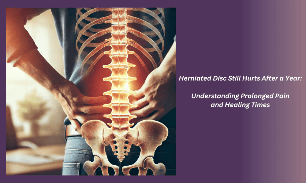
Quick Insights: Herniated Disc Pain Beyond One Year
Can a herniated disc heal after 2 years? Yes, but healing timelines vary significantly. While most herniated discs improve within 6-12 months, longitudinal studies show that disc herniation size continues to decrease over 2-7 years, though disc degeneration simultaneously progresses. The challenge: radiographic improvement doesn’t always correlate with symptom resolution.
Key finding: Persistent pain beyond one year often stems from annular tear repair failure and chronic inflammatory cascades from nucleus pulposus leakage—issues that conservative care cannot address. For patients whose herniated disc still hurts after a year despite appropriate treatment, interventional approaches targeting annular defects may offer the structural repair needed for lasting relief.
Introduction
For patients whose herniated disc still hurts after a year, understanding why pain persists becomes essential for finding effective relief. As an interventional spine specialist, I evaluate many patients in Houston who have followed conservative treatment protocols yet continue experiencing debilitating disc-related pain well beyond expected healing timelines.
When recovery extends beyond 12 months, specific questions emerge: “Can a herniated disc take 2 years to heal?” or “Why does my disc pain persist when imaging shows improvement?” These concerns reflect a critical reality in disc pathology—the disconnect between radiographic healing and symptomatic resolution. Longitudinal studies tracking herniated discs over 7+ years demonstrate that while herniation size decreases over time, disc degeneration progresses in all patients, and clinical symptoms don’t reliably predict long-term outcomes.
This article explores the mechanisms driving prolonged disc pain, evidence-based timelines for disc healing, and when structural disc repair—rather than symptom management—becomes medically appropriate. Whether you’re in the Houston, Katy, or Sugar Land area facing initial treatment delays or considering advanced interventional options, understanding the biology behind persistent disc pain can guide more informed treatment decisions.
Can a Herniated Disc Heal After 2 Years?
What Longitudinal Studies Reveal About Disc Healing Timelines
The natural history of herniated discs extends far beyond the 6-12 week timeframes often quoted in general practice. Research following patients with lumbar disc herniation for minimum 7 years shows that in nonsurgically treated patients, the space-occupying ratio of herniation diminishes progressively—with significant reduction by 2 years and continued improvement at final follow-up.
However, this radiographic improvement tells only part of the story. The same longitudinal data reveals that disc degeneration progressed in all patients during this extended timeline. More critically, clinical symptoms did not reliably predict long-term outcomes, highlighting the heterogeneity in healing patterns and the subset of patients whose herniated disc still hurts after a year despite imaging improvements.
A serial MRI follow-up study tracking herniated discs for an average of 30 months found that clinical improvement often occurs within the first year, and herniation size decreases over time. Yet the study emphasized that radiographic reduction does not always correlate perfectly with symptom resolution—a finding I observe regularly in clinical practice when evaluating patients whose pain persists despite “improved” MRI findings.
The Critical Difference: Herniation Resorption vs. Annular Repair
Understanding why some discs heal while others cause prolonged pain requires distinguishing between two separate processes:
Herniation resorption refers to the body’s immune response gradually breaking down and removing extruded disc material. Studies on natural history of extruded disc herniation show that extruded fragments demonstrate dynamic changes over time, with signal characteristics and size evolution relating to symptom duration. Larger extrusions with higher water content tend to shrink more predictably as inflammatory cells phagocytose the foreign tissue.
Annular repair, by contrast, addresses the tear in the disc’s outer wall (annulus fibrosus) through which the nucleus pulposus herniated. This distinction matters because herniation resorption may reduce nerve compression, but if the annular defect remains unhealed, nucleus pulposus material continues leaking into the epidural space, perpetuating inflammation and pain.
The annulus fibrosus has limited intrinsic healing capacity due to poor vascularization. When annular tears fail to seal, patients can experience persistent discogenic pain even as the herniation itself shrinks on imaging. This explains the clinical scenario where a patient’s MRI “looks better” at two years, yet their herniated disc still hurts—the structural defect driving pain was never addressed.
Can a Bulging Disc Heal After 2 Years?
Bulging discs follow different natural history patterns than frank herniations. A disc bulge represents circumferential or broad-based extension of disc material beyond vertebral margins without complete annular rupture. Because the annulus remains relatively intact in disc bulges, the acute inflammatory response from nucleus pulposus exposure is less pronounced than in full herniations.
However, disc bulges typically indicate underlying degenerative disc disease. The question “can a bulging disc heal after 2 years” depends on whether the disc’s degenerative process stabilizes or progresses. Serial MRI studies tracking conservatively treated discs for 30 months show that while some bulges reduce in size, disc degeneration advances regardless of treatment approach.
For bulging discs causing persistent symptoms beyond two years, the pain often reflects progressive disc collapse, facet joint overload, and biochemical inflammation rather than acute nerve compression. These cases may benefit from interventions targeting disc hydration and structural support rather than approaches designed for acute herniation management.

Why Your Herniated Disc Still Hurts After a Year
The Chronic Inflammatory Cascade
When a herniated disc still hurts after a year, chronic inflammation—not just mechanical nerve compression—often drives persistent pain. Research demonstrates that intervertebral discs causing low back pain secrete high levels of proinflammatory mediators, including interleukin-6 (IL-6), interleukin-8 (IL-8), and prostaglandin E2 (PGE2).
This inflammatory milieu creates a self-perpetuating pain cycle. As nucleus pulposus material continues leaking through unhealed annular tears, the immune system recognizes this tissue as foreign. Inflammatory cells infiltrate the area, releasing cytokines that sensitize nerve endings and maintain pain signaling—even when the original herniation has partially resorbed.
The biological mechanism explains why some patients report “good days and bad days” years after initial herniation. Activity that increases intradiscal pressure forces additional nucleus pulposus through the annular defect, triggering acute inflammatory flares. Anti-inflammatory medications may provide temporary relief by suppressing cytokine activity, but they cannot seal the structural pathway allowing continued nuclear material leakage.
Annular Tear Repair Failure
The annulus fibrosus consists of concentric collagen lamellae with minimal blood supply in its inner layers. When annular tears extend through multiple lamellae—as occurs in disc herniation—the body’s natural healing mechanisms face significant limitations.
Meta-analysis of annular repair outcomes reveals that annular closure techniques reduce recurrence and reoperation rates after discectomy compared with discectomy alone. This evidence supports what I observe clinically: addressing the annular defect matters as much as decompressing the nerve.
Without annular repair, the tear acts as a persistent pathway for nuclear material extrusion. Patients may experience temporary improvement as inflammation subsides, only to have symptoms return when physical activity increases intradiscal pressure. This pattern—improvement followed by recurrent pain—strongly suggests ongoing annular leakage rather than stable disc healing.
Factors Predicting Failed Conservative Treatment
Not all herniated discs respond equally to conservative care. Specific anatomical and patient factors predict which cases will have pain persisting beyond one year:
Herniation characteristics: Large extruded fragments with inferior migration demonstrate less predictable resorption patterns. Studies tracking extruded herniation natural history show that signal characteristics and hydration status influence how fragments evolve over time.
Duration before treatment initiation: Serial MRI follow-up data indicates that delayed intervention correlates with longer recovery timelines. Patients who wait months before starting appropriate therapy may allow chronic inflammatory changes and secondary muscle deconditioning to take hold.
Degree of disc degeneration: Severely degenerated discs lack the biomechanical stability and hydration needed to support natural healing. When the disc has lost significant height and the annular structure is globally compromised, conservative approaches cannot restore structural integrity.
Patient metabolic health: Diabetes, obesity, and smoking impair tissue healing through reduced blood flow and cellular dysfunction. These comorbidities extend recovery timelines and increase the likelihood that conservative treatment will prove insufficient.
Nerve Sensitization and Central Pain Processing
Prolonged peripheral nerve irritation from disc herniation can lead to neuroplastic changes in the central nervous system. When nerve roots experience sustained compression or chemical irritation from inflammatory disc material, the nervous system may become hypersensitized—a process called central sensitization.
In these cases, even after the mechanical compression resolves, the nervous system continues generating pain signals. Patients describe burning, electric-shock sensations, or hypersensitivity in the affected dermatome. This neuroplastic pain component explains why some individuals whose herniated disc still hurts after a year don’t experience proportional relief from interventions that successfully decompress the nerve.
Addressing central sensitization requires multimodal approaches that may include neuromodulation techniques alongside structural disc repair. The key insight: prolonged disc pathology creates both peripheral structural problems and central nervous system changes that must be recognized and treated appropriately.

When Conservative Treatment Stops Working
Recognizing the Plateau: Signs Conservative Care Has Reached Its Limit
Conservative treatment for herniated discs typically includes physical therapy, anti-inflammatory medications, activity modification, and sometimes epidural steroid injections. World Health Organization guidelines for non-surgical management of chronic primary low back pain provide evidence-based recommendations for these approaches, including when to escalate care.
However, conservative care has a therapeutic ceiling. I use specific clinical markers to identify when a herniated disc still hurts after a year because structural pathology—not inadequate symptom management—is driving persistent pain:
Pain plateau without functional gains: If pain scores remain unchanged after 6-9 months of appropriate conservative therapy, and functional limitations persist despite compliance with rehabilitation, the disc’s structural pathology likely exceeds what conservative measures can address.
Recurrent acute flares with minimal provocation: Patients who experience cyclical pain exacerbations—where minor activities trigger severe flares requiring days of bed rest—often have unhealed annular defects that permit nuclear material leakage with increased intradiscal pressure.
Progressive neurological findings: New or worsening weakness, sensory loss, or reflex changes despite conservative treatment indicate ongoing nerve compromise that requires structural intervention. While conservative care can manage symptoms, it cannot decompress a persistently compressed nerve root.
MRI-clinical discordance: When imaging shows persistent or worsening disc pathology while the patient reports “some improvement” from conservative care, this often indicates symptom suppression rather than structural healing. The disc’s underlying pathology remains unchanged.
The Role of Epidural Steroid Injections in Chronic Disc Pain
ASIPP comprehensive guidelines on epidural interventions provide evidence-based recommendations for these procedures in chronic spinal pain management. Epidural steroid injections can effectively reduce inflammatory-mediated pain and may facilitate participation in physical therapy for patients with acute radiculopathy.
However, epidural injections address inflammation, not structural disc pathology. When a patient receives multiple epidural injections with only temporary relief, this pattern suggests that inflammation is a secondary consequence of ongoing structural pathology rather than the primary problem. The injections suppress the inflammatory response, but as soon as the medication effect wanes, the unhealed annular tear continues producing inflammatory mediators.
I use response to epidural injections as a diagnostic tool: patients achieving 6-12 weeks of substantial relief likely have inflammation-predominant pain that may respond to continued conservative management. Those experiencing only 1-3 weeks of partial relief typically have structural disc pathology that requires more definitive intervention.
Understanding When “Watchful Waiting” Becomes “Waiting Too Long”
Natural history studies support conservative management for most acute herniated discs. Research on lumbar disc herniation treated without surgery shows that many herniations shrink over time, with imaging evolution correlating with symptom improvement in selected patients.
However, “watchful waiting” has time limits. When a herniated disc still hurts after a year despite appropriate conservative care, several concerning processes may be occurring:
Progressive disc degeneration: Longitudinal MRI data confirms that disc degeneration progresses during conservative treatment periods. Delayed intervention means treating more advanced pathology with potentially less favorable outcomes.
Secondary spinal changes: Chronic disc collapse alters spinal biomechanics, potentially accelerating adjacent segment degeneration and facet joint arthropathy. Early intervention to stabilize the affected disc may prevent these secondary problems.
Central sensitization development: Prolonged peripheral nerve irritation increases the risk of neuroplastic pain changes that persist even after structural problems are addressed. Earlier intervention may reduce this risk.
Functional deconditioning: Extended periods of pain-limited activity lead to muscle atrophy, cardiovascular deconditioning, and psychological distress that compound recovery challenges once treatment is initiated.
The decision to move beyond conservative care isn’t about treatment failure—it’s about recognizing when disc pathology exceeds what conservative approaches can structurally address.

The Discseel® Procedure: Addressing Annular Defects in Chronic Disc Pain
How Annular Repair Differs From Traditional Disc Surgery
Traditional surgical approaches to herniated discs focus on removing herniated material and decompressing nerves—either through microdiscectomy, endoscopic discectomy, or open decompression procedures. Long-term outcomes of standard discectomy show generally favorable results, though residual back pain and recurrent herniation occur in a meaningful subset of patients.
The limitation of traditional discectomy: while it removes the herniated fragment, it doesn’t seal the annular tear through which the herniation occurred. This leaves the pathway open for recurrent herniation and continued inflammatory mediator production from the disc interior.
Annular repair strategies take a different approach—sealing the defect to prevent ongoing nuclear material leakage. Meta-analysis of annular repair outcomes demonstrates that annular closure techniques reduce both recurrence and reoperation rates compared with discectomy alone.
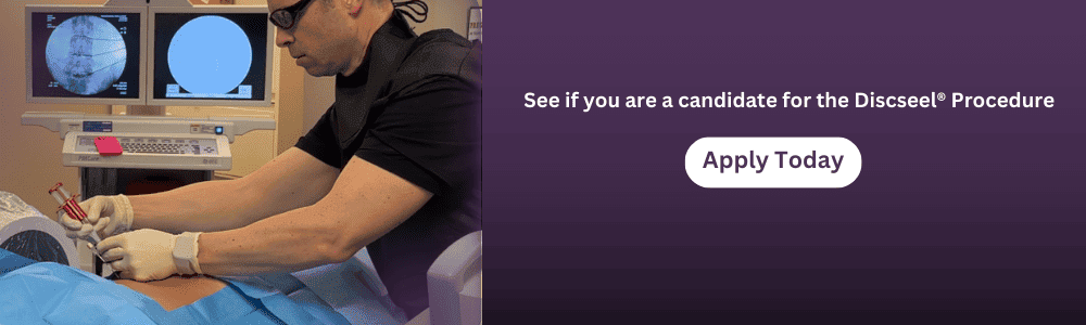
The Discseel® Procedure applies this annular repair concept through a minimally invasive, percutaneous approach. Rather than removing disc tissue through an open or endoscopic incision, Discseel® uses fluoroscopic guidance to deliver a biologic fibrin sealant directly into the disc and along annular defects.
The Science Behind Fibrin-Based Annular Sealing
Clinical studies on fibrin sealants in lumbar annuloplasty following endoscopic discectomy show favorable short-to-mid-term outcomes, with data suggesting reduced recurrence when fibrin is applied to annular tear sites. This evidence illustrates how biologic materials can facilitate annular healing in a clinical setting.
The Discseel® approach extends this concept by delivering fibrin throughout the disc’s interior and specifically targeting annular defects identified on discography imaging. The biologic sealant serves two functions:
Immediate mechanical closure: The fibrin matrix physically seals annular tears, creating a barrier that prevents ongoing nuclear material leakage into the epidural space. This immediate sealing may help reduce inflammatory mediator exposure to nerve roots.
Scaffold for collagen regeneration: Fibrin provides a three-dimensional scaffold that supports fibroblast infiltration and new collagen deposition. Over 3-6 months, the body’s natural healing mechanisms can generate new annular tissue within this scaffold, potentially achieving more durable structural repair than fibrin alone.
The procedure requires precise diagnostic annulargram imaging to identify actively leaking discs before treatment. Not every herniated disc demonstrates active leakage on provocation discography—Discseel® targets the subset where annular defects and ongoing nuclear extrusion drive persistent symptoms.
Patient Selection: When Discseel® Becomes Medically Appropriate
The Discseel® Procedure addresses specific disc pathology: annular tears with ongoing nuclear material leakage producing discogenic pain. I consider Discseel® evaluation appropriate when:
Conservative care has been optimized without adequate relief: The patient has completed 6-12 months of appropriate physical therapy, medication management, activity modification, and potentially epidural injections, yet continues experiencing functionally limiting pain.
Imaging confirms disc pathology consistent with symptoms: MRI demonstrates annular tears, disc degeneration, or herniation at levels corresponding to the patient’s pain distribution and examination findings.
The patient seeks to avoid or delay traditional spine surgery: Many patients whose herniated disc still hurts after a year face recommendations for fusion or disc replacement. Discseel® offers a minimally invasive alternative targeting disc repair rather than removal or replacement.
Pain is predominantly discogenic rather than facetogenic: Careful clinical examination and sometimes diagnostic injections help distinguish disc-driven pain from facet joint arthropathy or other spinal pain generators. Discseel® specifically targets disc pathology.
As the first physician in Houston trained directly by Dr. Kevin Pauza (who pioneered the Discseel® Procedure), I evaluate each patient’s specific anatomy, pain mechanisms, and treatment history to determine if this approach offers appropriate advantages over alternative interventions. The procedure isn’t suitable for all chronic disc pain—it addresses the subset where annular repair can resolve the structural problem perpetuating symptoms.
For patients in Houston, Katy, Sugar Land, or surrounding areas whose disc pain persists despite conservative treatment, Discseel® consultation can clarify whether this approach suits your specific pathology.
Other Advanced Treatment Options for Persistent Disc Pain
When Multiple Interventional Approaches May Be Needed
Some patients whose herniated disc still hurts after a year have complex pain generators requiring multimodal interventional strategies. Disc pathology rarely exists in isolation—facet joint arthropathy, sacroiliac joint dysfunction, and myofascial pain commonly coexist with chronic disc problems.
ASIPP guidelines on epidural interventions emphasize individualizing treatment based on specific pain mechanisms. This principle extends to all interventional spine care: accurate diagnosis of each pain generator guides appropriate procedure selection.
Regenerative Medicine Approaches Beyond Disc Sealing
Platelet-rich plasma (PRP) therapy represents another biologic approach to chronic disc pathology. PRP contains concentrated growth factors that may stimulate tissue repair and modulate inflammation. Some practitioners inject PRP into degenerative discs to promote healing, though the evidence base remains more limited than for annular repair techniques.
I view PRP as potentially complementary to—rather than replacing—annular repair strategies. PRP may optimize the healing environment, but it doesn’t mechanically seal annular defects the way fibrin-based approaches do. Patients considering PRP for disc pain should understand current evidence limitations and discuss realistic outcome expectations with their physician.
Spinal Cord Stimulation for Neuroplastic Pain Components
When chronic disc pathology has produced central sensitization and neuroplastic pain changes, spinal cord stimulation (SCS) may provide relief that structural disc interventions alone cannot achieve. SCS uses mild electrical signals to modulate pain processing in the spinal cord, potentially resetting hypersensitized neural pathways.
I consider SCS evaluation when patients report pain characteristics suggesting central sensitization—burning, electric sensations, allodynia (pain from normally non-painful stimuli), or pain distribution that doesn’t match anatomical dermatomes. These neuroplastic changes may require neuromodulation approaches alongside or instead of structural disc procedures.
The Role of Traditional Surgery: When It Remains the Best Option
Long-term discectomy outcome data confirms that traditional surgical decompression provides durable relief for appropriately selected patients. Despite the availability of minimally invasive alternatives, certain clinical scenarios still indicate traditional surgical approaches:
Progressive neurological deficits: New or worsening motor weakness, particularly affecting foot dorsiflexion or plantar flexion, requires urgent surgical decompression. Minimally invasive procedures cannot decompress large, sequestered fragments as reliably as direct surgical visualization.
Cauda equina syndrome: Saddle anesthesia, bladder/bowel dysfunction, or bilateral lower extremity weakness constitute surgical emergencies requiring immediate decompression. This represents a different clinical entity than chronic discogenic pain.
Failed prior interventions with persistent large herniation: When conservative care, injections, and potentially minimally invasive procedures have not provided adequate relief, and imaging shows substantial herniated material with ongoing nerve compression, surgical removal may be most appropriate.
The key is matching intervention to specific pathology. Patients whose herniated disc still hurts after a year have diverse underlying problems—some require annular repair, others need nerve decompression, and some benefit from neuromodulation. Comprehensive evaluation identifies which mechanisms drive each patient’s pain.

Prevention and Self-Management for Chronic Disc Pain
Spine-Protective Movement Patterns
Even while pursuing interventional treatment for a herniated disc that still hurts after a year, patients can adopt movement strategies that protect the spine and may prevent symptom exacerbations:
Hip-hinge mechanics for lifting: Bending from the hips rather than the lumbar spine reduces intradiscal pressure during lifting activities. This technique keeps the lumbar spine in neutral alignment, distributing load through the larger hip and leg muscles rather than concentrating stress on already-compromised discs.
Controlled rotation patterns: Combined flexion and rotation creates maximal shear stress on lumbar discs. When turning while lifting or reaching, patients should pivot with the feet rather than twisting through the spine, maintaining alignment between shoulders and hips.
Segmental stabilization: Engaging core muscles before and during activities that challenge the spine creates intrinsic spinal support. This isn’t about developing “six-pack abs”—it’s about coordinated activation of deep stabilizers (transversus abdominis, multifidus) that unload the disc.
Workstation Ergonomics for Disc Health
Prolonged sitting increases intradiscal pressure compared to standing or lying down. For patients working desk jobs while managing chronic disc pain, ergonomic modifications can reduce mechanical stress:
Lumbar support positioning: A small roll or cushion supporting the lumbar lordosis helps maintain the spine’s natural curves during sitting. This external support reduces the muscular effort needed to maintain posture, potentially decreasing fatigue-related pain.
Monitor and keyboard placement: Screen height at eye level and keyboard position allowing relaxed shoulder posture prevent compensatory postural adaptations that increase spinal loading. Neutral spine positioning during computer work reduces cumulative disc stress over an 8-hour workday.
Sit-stand workstation strategies: Alternating between sitting and standing positions throughout the day varies spinal loading patterns and prevents the disc hydration changes that occur with prolonged static postures. I recommend position changes every 30-45 minutes rather than extended periods in either position.
Activity Modification Without Complete Activity Restriction
A common error in managing herniated disc pain: excessive activity restriction leading to deconditioning. WHO guidelines for chronic low back pain management emphasize maintaining appropriate activity levels rather than prolonged bed rest or complete activity avoidance.
The goal is pain-guided progression—engaging in activities up to moderate discomfort levels without provoking severe flares. Walking programs, aquatic therapy, and graduated resistance training can maintain conditioning even while disc pathology is being addressed through interventional means.
For patients whose herniated disc still hurts after a year, this balance proves challenging. Severe pain naturally limits activity, yet excessive deconditioning impairs recovery once effective treatment is initiated. Working with physical therapists experienced in chronic spine conditions helps navigate this challenge.
Weight Management and Metabolic Health
Excess body weight increases mechanical loading on lumbar discs, particularly at the lumbosacral junction where most herniations occur. Additionally, adipose tissue produces inflammatory cytokines that may contribute to disc degeneration and pain amplification.
Weight reduction, when medically appropriate, can reduce disc loading and systemic inflammation. However, I emphasize that weight loss alone rarely resolves established disc pathology—it’s a supportive measure rather than primary treatment for herniated discs. Patients shouldn’t delay evaluation and treatment while pursuing weight loss goals, as structural disc problems typically require specific interventions regardless of weight management efforts.
Metabolic conditions like diabetes impair tissue healing and may affect outcomes from both conservative and interventional disc treatments. Optimizing glucose control, addressing nutritional deficiencies, and managing cardiovascular risk factors support the body’s healing capacity once appropriate disc treatment is initiated.
Conclusion
When a herniated disc still hurts after a year, this timeline indicates more than slow healing—it often reflects structural disc pathology that conservative care cannot adequately address. Longitudinal studies tracking herniated discs over 2-7 years demonstrate that while herniation size may decrease over time, disc degeneration progresses, and symptom resolution doesn’t reliably correlate with imaging improvements.
The critical insights: persistent disc pain beyond 12 months frequently stems from unhealed annular defects that permit ongoing nuclear material leakage and inflammatory mediator production. Research confirms that painful discs secrete high levels of proinflammatory cytokines, creating a biochemical environment that perpetuates pain independent of mechanical nerve compression.
For Houston-area patients whose conservative treatment has reached its therapeutic ceiling, understanding these mechanisms clarifies why symptoms persist and which interventions may offer more definitive relief. Evidence showing that annular repair techniques reduce recurrence and reoperation rates supports the biological rationale for procedures like Discseel® that target structural disc defects rather than just managing symptoms.
The question “can a herniated disc heal after 2 years” has a nuanced answer: yes, continued herniation resorption occurs over extended timelines, but true healing requires both herniation resolution and annular repair. When annular defects remain unhealed, patients experience the frustrating cycle of improvement and recurrent flares that characterizes chronic discogenic pain.
If your herniated disc pain persists despite appropriate conservative care, consultation with an interventional spine specialist can clarify whether your specific pathology exceeds what conservative approaches can structurally address. At Performance Pain and Sports Medicine in Houston, we provide comprehensive evaluation to identify the specific mechanisms driving your pain and determine which interventions—from continued conservative care to advanced procedures like Discseel®—offer the best opportunity for lasting relief.
Schedule a consultation to discuss whether your chronic disc pain would benefit from interventional evaluation. Understanding the structural basis for prolonged disc pain is the first step toward finding effective treatment.
Frequently Asked Questions
How long does it take for a herniated disc to fully heal?
Most herniated discs show significant improvement within 6-12 months, with herniation size progressively decreasing over 2-7 years according to longitudinal MRI studies. However, “healing” involves two distinct processes: herniation resorption (the body breaking down extruded disc material) and annular repair (sealing the tear through which the disc herniated). While herniation resorption follows predictable timelines, annular defects may never fully heal without intervention due to the annulus fibrosus’s limited blood supply and intrinsic repair capacity.
Why does my MRI look better but I still have pain?
This common scenario—improved imaging with persistent symptoms—typically indicates that while the herniated fragment has partially resorbed, the underlying annular tear remains unhealed. Unhealed annular defects allow continued leakage of nucleus pulposus material into the epidural space, producing inflammatory cytokines that maintain pain even without significant nerve compression visible on MRI. This imaging-clinical discordance suggests discogenic pain from annular pathology rather than pure nerve compression pain.
Can a herniated disc get worse after a year?
Yes, herniated discs can progress even after the initial injury. Re-injury from premature return to high-impact activities, progressive disc degeneration weakening adjacent annular layers, or enlargement of the original annular tear can all worsen disc pathology over time. Additionally, some patients develop secondary problems like adjacent segment degeneration or facet joint arthropathy that compound the original disc pain. Worsening symptoms after initial improvement warrant re-evaluation to identify whether the original disc pathology has progressed or new problems have developed.
What percentage of herniated discs require surgery?
Approximately 10-20% of patients with herniated discs ultimately undergo surgery, though this varies based on patient population and surgical indications. Most herniated discs respond adequately to conservative care within 6-12 months. However, certain factors increase the likelihood of surgical intervention: large extruded fragments with inferior migration, progressive neurological deficits, cauda equina syndrome, or persistent functionally limiting pain despite 6-12 months of appropriate conservative treatment. The decision for surgery should be individualized based on specific pathology, symptom severity, and patient treatment goals.
Is the Discseel® Procedure the same as traditional spine surgery?
No, Discseel® differs fundamentally from traditional spine surgery in both approach and mechanism. Traditional discectomy removes herniated disc material through open or endoscopic incision to decompress nerves but doesn’t seal the annular defect. Discseel® uses fluoroscopic-guided needle access to deliver fibrin sealant directly into the disc and along annular tears, sealing defects percutaneously without removing tissue. The procedures address different aspects of disc pathology: traditional surgery focuses on decompression, while Discseel® targets annular repair and disc stabilization.
When should I consider interventional treatment instead of continuing conservative care?
Consider interventional evaluation when: (1) pain persists beyond 6-12 months despite optimized physical therapy, medications, and activity modification; (2) you experience cyclical pain flares with minimal provocation, suggesting unhealed annular defects; (3) epidural injections provide only temporary (1-3 weeks) relief rather than sustained improvement; (4) functional limitations prevent work or essential daily activities despite conservative treatment compliance; or (5) you face recommendations for fusion or disc replacement but want to explore less invasive alternatives first. Interventional consultation doesn’t commit you to procedures—it clarifies whether your specific pathology exceeds what conservative care can structurally address.
This article is for educational purposes only and should not be used as a substitute for professional medical advice, diagnosis, or treatment. Always seek the advice of your physician or other qualified healthcare provider with any questions you may have regarding a medical condition or treatment options. Never disregard professional medical advice or delay in seeking it because of something you have read in this article.