By Dr. Matthias Wiederholz, MD
Quadruple Board-Certified in Physical Medicine & Rehabilitation, Sports Medicine, Pain Medicine, and Anti-Aging, Regenerative & Functional Medicine
Quick Insights
Learning how to read an MRI for a herniated disc helps you understand what your imaging shows. MRI uses magnetic fields to create detailed pictures of your spinal discs, nerves, and surrounding tissues. The scan reveals disc height, tears, herniations, and nerve compression. However, MRI findings don’t always match pain levels—many people have disc abnormalities without symptoms. Understanding your MRI empowers you to ask better questions and make informed treatment decisions with your physician.
Key Takeaways
- Annular tears and disc protrusions appear in 56% of people without back pain or sciatica.
- Foraminal narrowing and nerve root compression on MRI correlate most strongly with radicular symptoms.
- Pfirrmann grading shows disc degeneration severity but has limited value for predicting symptom acuity.
- Advanced MRI techniques like diffusion tensor imaging can reveal nerve inflammation not visible on standard scans.
Why It Matters
Understanding how to read an MRI for a herniated disc transforms confusing medical reports into actionable knowledge. This clarity helps you evaluate treatment recommendations confidently, avoid unnecessary procedures, and recognize when imaging findings require expert interpretation. When you understand what your MRI shows—and what it doesn’t—you can participate actively in decisions about your spine health and long-term mobility.
Introduction
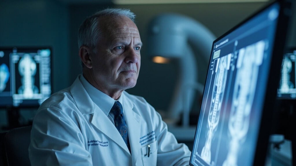
As a quadruple board-certified physician serving Houston and specializing in spine care, I’ve reviewed thousands of MRI reports with patients who feel overwhelmed by confusing terminology. Terms like “annular tear,” “disc protrusion,” and “foraminal stenosis” can seem intimidating when you’re trying to understand what’s causing your pain. Learning how to read an MRI for a herniated disc empowers you to have informed conversations with your physician and make confident treatment decisions.
MRI uses magnetic fields to create detailed images of your spinal discs, nerves, and surrounding tissues. These scans reveal disc height, structural tears, herniations, and nerve compression patterns. However, research shows that disc degeneration visible on MRI doesn’t always correlate with pain severity—many people have significant disc abnormalities without any symptoms. Understanding this gap between imaging findings and clinical symptoms is crucial for evaluating treatment recommendations.
At Performance Pain and Sports Medicine, I help patients from Montgomery to Shenandoah translate complex radiology reports into actionable knowledge. When you understand what your MRI shows—and what it doesn’t predict—you can participate actively in decisions about your spine health and avoid unnecessary procedures. For a deeper look at spinal disc tears and what your imaging reveals, see The Guide to Spinal Disc Tears: Causes, Symptoms, and Treatment Options. I also welcome patients seeking more information about my expertise to visit my physician bio.
If you’re dealing with ongoing back discomfort, learning about effective treatment options for L5-S1 disc herniation pain may help guide your next steps.
What Does an MRI Show?
MRI uses powerful magnetic fields to create detailed images of your spine’s soft tissues. Unlike X-rays that only show bones, MRI reveals discs, nerves, spinal cord, and surrounding structures. The scan shows disc height, hydration levels, tears in the disc wall, and any material pressing on nerves.
In my Houston practice, I use MRI to identify the specific disc level causing problems and assess structural damage. Research confirms that MRI helps determine herniation type, size, and location, which guides treatment decisions. The images show whether disc material has moved beyond normal boundaries and if nerves are compressed.
However, MRI cannot measure pain intensity or predict how you’ll respond to treatment. The scan captures a single moment in time and doesn’t show inflammation levels or nerve irritation that may be causing your symptoms. This is why I always correlate MRI findings with your physical exam and symptom pattern. For a comprehensive understanding of back pain and its relationship to MRI findings, you can explore our extensive resources.
If you’re interested in types of disc abnormalities, such as a bulging disc in the neck region, these conditions may also be visualized on MRI and help further explain your symptoms.
Common MRI Terms for Herniated Discs Explained
Understanding radiology terminology helps you interpret your report. A disc bulge occurs when the disc extends beyond the vertebra. A protrusion occurs when disc material extends beyond the normal confines of the disc space without complete disruption of the annulus fibrosus. An extrusion occurs when disc material extends beyond the disc space with complete disruption of the annulus fibrosus and may separate from the disc.
Annular tears or fissures are cracks that may develop in the outer disc wall. These tears may allow inner disc material to leak out, potentially triggering inflammation. Disc desiccation refers to the loss of water content in the disc, which may appear darker on MRI. Pfirrmann grading scores disc degeneration from 1 (healthy) to 5 (severely degenerated).
Foraminal stenosis describes the narrowing of the openings where nerve roots exit your spine. Studies show that foraminal compromise and specific herniation patterns correlate most strongly with radicular symptoms like leg pain or arm pain. Nerve root impingement means disc material or bone is pressing directly on a nerve.
Want to know more about disc damage and regeneration? See our resource on disc desiccation, including causes, symptoms, and treatment.

How to Identify a Herniated Disc on MRI Images
MRI scans use different views to show disc problems. Sagittal images show your spine from the side, revealing disc height and alignment. Axial images are cross-sections that show nerve compression and disc material extending into the spinal canal.
Healthy discs typically appear bright on T2-weighted images due to their water content. Degenerated discs may appear darker on T2-weighted images. Herniated disc material appears as a bulge or protrusion extending beyond the normal disc boundary.
Research demonstrates that degenerative changes and herniation patterns are extremely common on MRI, even in people without symptoms. When I review your images, I look for specific features that explain your pain pattern—not just any abnormality. The location and size of a herniation matter, but so does whether it’s compressing a nerve root.
If you’d like a step-by-step breakdown of interpreting herniated disc MRI lumbar scans, check our detailed guides.

What MRI Findings Mean for Your Symptoms
Not all MRI findings cause symptoms. Central disc bulges often don’t produce pain unless they’re large enough to compress the spinal cord. Foraminal herniations that narrow nerve exit pathways typically correlate with radiating arm or leg pain. Direct nerve root compression usually causes specific dermatomal symptoms.
In my Houston-area practice, I focus on findings that match your clinical presentation. If you have right leg pain, I look for right-sided nerve compression at the appropriate level. Advanced MRI techniques like diffusion tensor imaging can reveal nerve inflammation that explains symptoms when standard images look relatively normal.
Annular tears visible on MRI may indicate discogenic pain—pain originating from the disc itself rather than nerve compression. These tears allow inflammatory proteins to leak out and irritate pain-sensitive nerve fibers in the outer disc wall. This mechanism explains why some patients have severe back pain without obvious nerve compression on MRI.
Explore Disc Tear Treatment: Restoring the Integrity of Your Spine for more on advanced care options for MRI-identified tears.
The Gap Between MRI Results and Pain Levels
One of the most important concepts I teach patients is that MRI severity doesn’t predict pain severity. Studies confirm that MRI features have limited predictive value for symptom acuity and intensity. You can have a dramatic-looking herniation with minimal pain, or severe pain with subtle MRI changes.
Landmark research found that annular tears and disc protrusions appear in 56% of people without back pain or sciatica. This means many MRI abnormalities are incidental findings that don’t require treatment. Age-related disc changes are normal and don’t automatically cause symptoms.
This gap between imaging and symptoms is why I never recommend treatment based solely on MRI findings. Your pain pattern, physical exam, and response to conservative care matter more than how your MRI looks. Some patients need intervention for relatively mild MRI findings because those specific changes are causing their symptoms, while others with worse-looking scans improve with conservative treatment.
For further clarity on herniated disc symptoms, causes, and treatment, consult additional guides to distinguish imaging from clinical realities.

One Patient’s Experience
As a physician who reviews MRI reports daily, I understand how confusing imaging findings can feel when you’re trying to decide on treatment.
I had herniations on all my lumbar discs on both sides of my spine and also on the first sacral disc. I couldn’t walk without leaning on something and was in constant pain. Went to a spine surgeon who wanted to fuse my bottom lumbar disc to my sacral disc. When I asked him about the pain farther up the lumbar spine, he just said that was referred pain. Dr. Wiederholz took the time to review my entire MRI and explained how multiple disc levels were contributing to my symptoms. His comprehensive approach with the Discseel® Procedure addressed all the affected areas my previous surgeon had dismissed. Now I can walk normally, travel to the Virgin Islands, and enjoy activities I thought I’d lost forever. It’s been 20 years since I felt this good.
Read the full patient experience on Google
This is one patient’s experience; individual results may vary.
Linda’s MRI showed multiple lumbar herniations, but her previous surgeon focused only on one level. Expert MRI interpretation means evaluating all affected discs and correlating findings with your complete symptom pattern, not dismissing pain as “referred” when imaging shows structural problems at multiple levels.
Want to see how Discseel® reviews reflect lasting back pain relief? Check more patient stories of restored function.
Conclusion
Learning how to read an MRI for a herniated disc empowers you to understand what your imaging reveals about disc structure, nerve compression, and degenerative changes. However, MRI findings must always be interpreted by an experienced physician who correlates imaging with your symptoms, physical examination, and treatment history.
Many disc abnormalities visible on MRI occur in people without pain, while some patients experience severe symptoms with subtle imaging changes. This gap underscores why expert clinical correlation is essential—not all MRI findings require treatment, and not all pain has dramatic MRI features.
As a quadruple board-certified physician specializing in disc-focused diagnostics in Houston, I help patients understand when MRI findings indicate structural disc pathology that may benefit from advanced interventions. When conservative care has been insufficient, and your MRI reveals annular tears or disc damage contributing to persistent symptoms, comprehensive evaluation by a spine specialist can determine whether mechanism-based options like the Discseel® Procedure may be appropriate.
Understanding your MRI is the first step toward informed treatment decisions. Whether you’re in Oak Ridge North or the surrounding areas, if you’re ready to have your imaging interpreted by a physician who specializes in disc pathology at Performance Pain and Sports Medicine, see if you may be a candidate for the Discseel® Procedure.
Looking for treatment options for degenerative disc disease or exploring care at a Houston-area facility? Learn more about our comprehensive, locally-focused spine care.
This article is for educational purposes only and should not be used as a substitute for professional medical advice, diagnosis, or treatment. Always seek the advice of your physician or other qualified healthcare provider with any questions you may have regarding a medical condition or treatment options. Never disregard professional medical advice or delay in seeking it because of something you have read in this article.
Frequently Asked Questions
Can an MRI show a healed herniated disc?
MRI can show changes in disc appearance over time, including a reduction in herniation size or rehydration of previously desiccated discs. However, MRI cannot definitively determine if a disc has “healed” in the sense of restored structural integrity. Annular tears may remain visible even after symptoms improve, and disc material that has extruded may be resorbed by the body. The key question isn’t whether your MRI looks better, but whether your symptoms have resolved and function has been restored through appropriate treatment.
If you want to understand the process of healing and recovery, explore the differences in MRI findings between normal and herniated discs.
Does a worse-looking MRI mean I’ll have more pain?
No. MRI severity does not predict pain intensity. Research shows that many people with dramatic-looking herniations, multiple-level degeneration, or large disc protrusions have minimal or no symptoms, while others experience severe pain with relatively mild MRI findings. Pain depends on factors MRI cannot measure—nerve inflammation levels, central sensitization, muscle dysfunction, and individual pain processing. This is why I always correlate your imaging with your symptom pattern and physical examination rather than treating the MRI alone.
For additional insights into imaging and symptom disconnect, review emergency symptoms of a herniated disc and when imaging matters most.
What MRI findings indicate I might need advanced intervention?
Appropriateness criteria emphasize that MRI findings should guide intervention only when correlated with persistent symptoms despite conservative care. Findings that may warrant advanced evaluation include foraminal stenosis with corresponding radicular symptoms, annular tears with discogenic pain patterns, or progressive neurological deficits with nerve compression.
However, the decision for any intervention depends on your complete clinical picture—including symptom duration, functional impact, conservative treatment response, and overall health—not imaging appearance alone.
You can learn more about ruptured disc MRI findings and next steps if your report suggests severe disc pathology.
Where can I find expert MRI interpretation for herniated discs in Houston?
At Performance Pain and Sports Medicine in Houston, Dr. Matthias Wiederholz provides comprehensive MRI interpretation for patients with herniated discs. As a quadruple board-certified physician and Master Instructor for the Discseel® Procedure, Dr. Wiederholz specializes in correlating imaging findings with clinical symptoms to determine the most appropriate treatment approach.
He serves patients throughout the Houston area, including those from nearby communities like Houston Methodist, The Woodlands Hospital, and Memorial Hermann The Woodlands. Expert interpretation goes beyond reading the radiology report—it involves understanding which findings explain your specific symptoms and when advanced interventions may be appropriate after conservative care. If you’re ready to start your journey to relief, contact us today.